 The Digestive Functions Of
The Human Body
The Digestive Functions Of
The Human Body
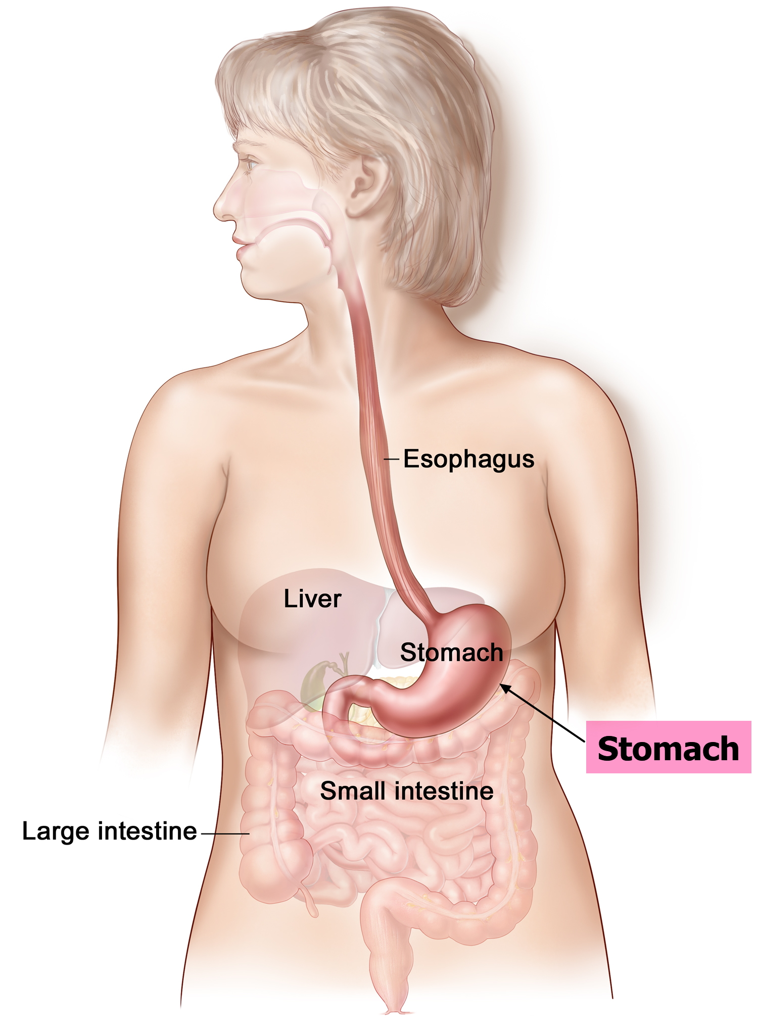
The Oesophagus is a long muscular tube lined up with mucous membrane, which runs behind the windpipe,(trachea), heart and in front of the spine. The Oesophagus connects to the throat and stomach. When you have chewed the food, using saliva, it lubricates the food turning it into a bolus, ( Latin word for "ball"), so it can be digested easily. Then the walls in the Oesophagus squeeze together, moving the bolus down to the stomach to be digested. This process is called Peristalsis. The esophagus is important because, it is an essential piece of the gastrointestinal tract, and functions as a pipe, for food and liquids that have been swallowed.
For Example In a Giraffe :
Peristalsis operates through the movement of longitudinal and circular muscles, along the walls of the esophagus, stomach, and intestine. A giraffes esophagus is approximately 1.8 meters long and weighs 272 kilograms. Also, like cows giraffes have four stomachs which indicate they are ruminants. The four stomachs assist with digesting food.
 The Human Stomach:
The stomach is a muscular organ positioned on the left-hand side of the upper abdomen. The human stomach is similar to a large elastic bag, which can expand to hold up to 2 to 4 litres of food. The stomach is connected to the esophagus and duodenum. The stomach produces acids ( Pepsin, Hydrochloric, and Mucus ) and enzymes that digest food. Then muscle tissue called rugae, line the stomach up, and next, the stomach muscles decrease and increase rhythmically, mixing food with the acid and enzymes to start breaking down the food. The food ends up as a paste called Chyme. After that, the pyloric sphincter which is a muscular valve opens and allows chyme to pass through to the small intestine. The stomach is important because it digests food using acids and enzymes, without it, we would not be able to digest food. Therefore nutrients would not be able to enter the bloodstream. The stomach has an important role in the immune system, which helps fight infections. Did you know, you can live without a stomach. The main function of the stomach is to collect and break down food, so it can slowly move through into the small intestine. Therefore the body is capable of surviving without a stomach. In result, food must be eaten in small portions so it can proceed straight to the small intestine.
For Example A Cow :
A cow has four stomachs the Rumen, Reticulum, Omasum, and Abomasum. The four stomachs share a special digestive process, to break down tough and coarse foods it consumes. When the cow first eats, it chews just barely to swallow it. The unchewed food of the cow then proceeds to the first two stomachs, the rumen, and the reticulum. Next, the cow will cough up pieces of the unchewed food called cud, and later chews it completely and then swallows it. The cud then moves to the third and fourth stomach ( Omasum & Abomasum ) to be completely digested.

The Small Intestine :
The small intestine is a long and convoluted tube, that is responsible for the absorption of nutrients from the food we eat. The small intestine is the section where 90% of the digestion and absorption of food happens. The remaining 10% happens in the stomach and large intestine. It lies between the stomach and the large intestine ( colon ). The small intestine has three major sections the Duodenum, Jejunum & the Ileum. The Duodenums is the first region which connects to the stomach. Partly digested food or chyme, from the stomach, is combined with bile from the liver, and pancreatic liquid from the pancreas. Which concludes its digestion in the duodenum. Next, we have the Jejunum, in the medial section, which plays the primary section of absorption. Lastly, we have Ileum which completes the absorption of the nutrients which was missed by the Jejunum. The interior walls of the small intestine are surrounded with finger-like projections called villi. The surface of the villi is covered with more, smaller finger-like projection called microvilli. They are there to improve the rate of absorption. Chyme is slowly moved through the small intestine by waves and muscles contraction called Peristalsis. Peristalsis starts at the duodenum, then the Jejunum and lastly Ileum. The small intestines main function is to absorb the nutrients and minerals from food and then deliver it into our bloodstream. Without the small intestine, the food we eat won't be able to enter our bloodstream. The small intestine is longer than the large intestine but the large intestine is wider than the small intestine.
For Example A Elephant:
An intestine of an elephant can spread up to 19 meters in length. The intestine is where most of the digestion of the vegetative diet happens. Elephants only use 40% of their intake. The size of an elephant's feces is based on their age, but an adult African elephant can excrete up to 300 pounds ( 136 kg ) of feces each day. Elephants absorb so few nutrients in result their feces are rich solid food matter and nutrients. Therefore it is advantageous for other animals.
The Large Intestine ( Colon ) :
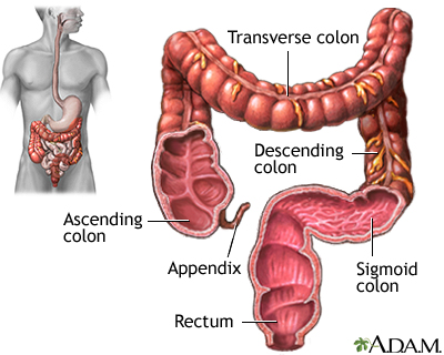
The large intestine is the final segment of the gastronomical tract and can also be called colon. The large intestine is a long and convoluted tube running from the small intestine to the anus. It surrounds the small intestine. It absorbs water from the undigestible food while, turning digested food into feces and excretes it out of the body. The cecum is the beginning of the large intestine and is connected to the last part of the small intestine the Ileum. The cecum gives a mixing space for bacteria with inadequately digested food, from the small intestine to develop feces. Then comes the ascending colon. One of the four significant colons in the large intestine, the ascending colon carries feces from the cecum to the transverse colon. The inner walls of the ascending colon absorb water and nutrients from the feces. Then comes the hepatic flexure which connects to the ascending colon and the transverse colon. The transverse colon is up next and many of the absorptions and formation of feces happen here making it a very significant colon. Following the transverse colon, the splenic flexure is next and connects to the transverse colon and descending colon. Then comes the descending colon another important colon out of the four, this colon stores feces until they are ready for them to be excreted from the body. The walls of the descending colon collect the remaining nutrients and absorb all the water. Lastly comes the sigmoid colon which transfers feces from the descending colon to the rectum. This colon also stores feces and releases them when they are ready. After that comes the rectum and anus. Why do we need the large intestine? Well without the large intestine the chyme from the stomach would not be able to absorb water and nutrients turning them into feces. The large intestine is an important part of the gastronomical tract. Did you know you can live without a large intestine, but you have to wear a bag outside your body in order to collect stool.
The large intestine in a camel is about 78 feet long ( 23.8 metres ). Half of the large intestine's job is to absorb water, salts ( sodium chloride ) and vitamins produced by bacteria. The other half's job is to store the feces in its large intestine until it excretion time.
The Liver :
The liver is the second largest organ in the human body, following the skin. The liver is positioned on the upper right quadrant of the abdomen, below the diaphragm and guarded by the lower ribs. The liver is a large organ in the abdomen in vertebrates. The liver has two large parts called the left lobe and the right lobe. The liver's main job is to filter blood coming from the digestive tract before passing it into the rest of the body. Another function is after food from the small intestine is digested, the nutrients are absorbed and carried to the liver within the hepatic portal. These nutrients ( glucose, amino acids, fatty acids and glycerol ) are either transformed into energy, moved to work muscles or saved for later use. Another important function of the liver is it produces a green liquid called bile. Bile is collected in the gallbladder and transported to the duodenum through the bile duct. Why do we need the liver? Well without the liver we would not be able clean blood, produce an important liquid called bile or store energy. The liver is very important since produces proteins which are significant in blood clotting, with the help of vitamin K.
For Example in a Dog:
A dogs liver is complicated and hard working. The liver in a dogs body is the biggest organ. The liver has many functions in dogs here are some: The making of bile, which is delivered into the gastrointestinal tract to help break down fats in the small intestine during digestion, the production of cholesterol and specific proteins that help transport fats throughout the body and the creation of certain proteins that circulate in the blood. A dogs liver is almost similar to humans liver.
-------------------------------------------------------------------------------------------------------------
Later last week in science class we were given a lamb to dissect. Unfortunately, the lamb didn't live through winter hence why we got to dissect it. While I was looking at the lamb's digestive system I found it was very similar to the human digestive system. The positioning of the gastronomical tract seemed similar to humans. We were allowed to touch the lamb's body parts and the feeling of the lamb's oesophagus felt very comparable to rubber. Something I found cool or interesting was the touch of the intestines it felt like thick noodles. Overall I enjoyed learning about the lamb's digestive system and the food and digestion section in the scipad. Sadly, I was able to take a photo.
----------------------------------------------------------------------------------------------------------------------------------
Bibliography :
Stomach
Small Intestine
Oesophagus
Large Intestine
Liver

 Preparing A Slide ( Microscope )


 Aim: To test if a sample of food contains protein.
 TESTING FOR SIMPLE SUGARS - THE BENEDICTS TEST:
 TESTING FOR COMPLEX SUGARS - THE IODINE TEST :


 Features:
 Features:
 Features:
 Features:
 Features:
 Features:
 Features:
 Features:
  Features:
 Features: